Also known as: Exercise echo or stress echo
Duration: About 60 minutes
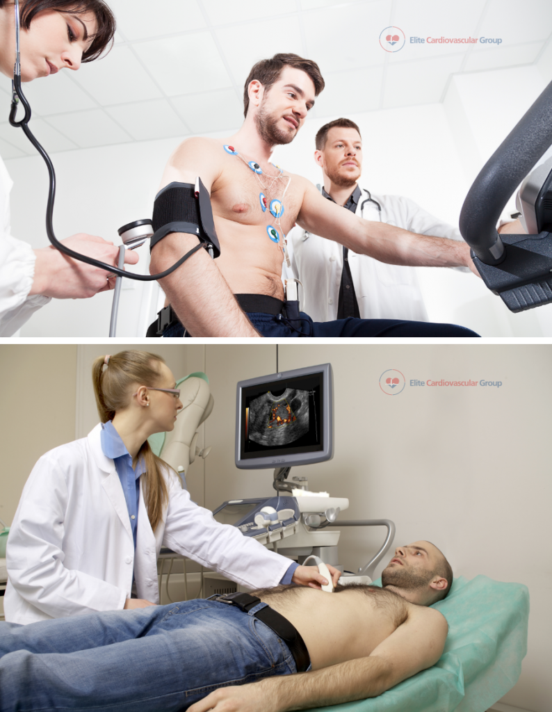
The stress echo is the combination of an exercise stress test with an echocardiogram. It is a noninvasive test that is used to compare how the heart performs under “stress”/exercise to how the heart is performing at rest.

Uses:
- To measure blood pressure during exercise
- To check the heart chambers, the major blood vessels, the thickness of the heart muscle, and the function of the valves
- To check for coronary artery disease (CAD)
- To check how well the heart is pumping
- To make sure that exercise can be tolerated by someone recovering from heart surgery, a heart attack, or someone going into cardiac rehabilitation
Preparing for the test:
![]() Download Pre Test Instructions
Download Pre Test Instructions
How it is performed:
- Echo imaging is performed before and after the exercise stress test.
- After the exercise portion is completed, you may be asked to stand still and then lie down for some time to check your heart’s recovery.
- The echo machine takes pictures of the heart by using an ultrasound probe/transducer. The probe sends out sound waves to the heart and blood vessels and an image is created when it picks up sound waves that bounce back from the heart and blood vessels.
- Once completed, your cardiologist will compare the results for both the resting echo and the stress echo.
After the test:
- Your cardiologist will discuss the results and the next steps of action.
- You can continue your daily activities (unless told otherwise).
- Ask your cardiologist if you have any questions or concerns.
Show references
Steeds RP, Wheeler R, Bhattacharyya S, et al. Stress echocardiography in coronary artery disease: a practical guideline from the British Society of Echocardiography. Echo Res Pract. 2019;6(2):G17-G33. doi:10.1530/ERP-18-0068
Suzuki K, Hirano Y, Yamada H, et al. Practical guidance for the implementation of stress echocardiography. J Echocardiogr. 2018;16(3):105-129. doi:10.1007/s12574-018-0382-8
Sicari R, Cortigiani L. The clinical use of stress echocardiography in ischemic heart disease. Cardiovasc Ultrasound. 2017;15(1):7. Published 2017 Mar 21. doi:10.1186/s12947-017-0099-2
