Also known as: Transfemoral venoplasty or transfemoral vein angioplasty
Duration: 30 minutes (but can take longer depending on complexities)
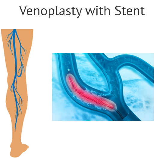
Peripheral venoplasty is a minimally invasive procedure used to open up blocked or narrowed leg veins. It follows the peripheral venogram and restores blood flow to the lower extremities/limbs without the need for vascular surgery. This procedure is usually followed by placing a stent (a short wire-mesh tube) into the affected vein.
Uses:
- For persistent and acute leg pain and swelling
- For chronic venous insufficiency and varicose veins
- For deep venous thrombosis (DVT)
Preparing for the procedure:
![]() Download Pre Procedure Instructions
Download Pre Procedure Instructions
How it is performed:
- Firstly, a peripheral venogram is performed to check for any blocks in the arteries of the legs. A catheter (a thin, long tube) is placed in either your arms, legs, or groin and is monitored by an x-ray camera until it reaches the targeted artery (arteries).
- A contrast dye is then infused so that the arteries can be seen.
- If a vein is blocked, you may require a balloon angioplasty to open it up.
- In balloon angioplasty, the tip of the catheter will be fitted into the blocked vein and the balloon is inflated. This pushes the plaque against the wall of the artery, creating more space for blood to flow easily.
- Once blood flow is established, the catheter is deflated and removed.
Stent Placement
- Most patients who undergo balloon angioplasty may also have a stent (a small, collapsed tube made of wire mesh) placed inside the treated vein.
- The balloon of the stent is inflated, causing it to expand against the wall of the vein.
- Once placed, the balloon is deflated and withdrawn.
Types of Stents:
- Drug-Eluting Stents (DES) – is coated with medication to keep the veins from clogging again. Most stents are of this type.
- Bare Metal Stents (BMS) – is not coated with medication
After the procedure:
![]() Download Post Procedure Instructions
Download Post Procedure Instructions
Show references
Jackson KP. Venoplasty and Stenting. Card Electrophysiol Clin. 2018;10(4):675-680. doi:10.1016/j.ccep.2018.07.005